Up Close & Personal: Anatomy Class Visits Body Worlds Exhibit
Angus Fung was one of the 30 seniors from the Anatomy and Physiology class who attended a field trip on Friday, April 24, 2015. He saw guts, brains, reproductive organs, fetuses, and other grotesque body parts. Although such images may cause the average teenager to get sick, these students believed that they were part of an interesting experience at the Body Worlds exhibit in New York City.
“I think it was definitely worth it to see the tangible and physical aspects that we otherwise only see in textbooks on a day-to-day basis,” Fung said.
A controversial exhibit at Discovery Times Square, “Body Worlds: Pulse” allows visitors to view anatomical structures preserved through plastination, a technique invented by German scientist Gunther von Hagens in 1975. Von Hagens, the founder of the Body Worlds exhibits, received much criticism for his disrespectful displays because many Catholics believed that dead bodies should be buried, not shown in different positions for the people’s enjoyment. There were even allegations that none of the bodies were shown with consent. However, Hagens insisted that all of the bodies and parts were donated by people who gave consent.
“If you’re an adult and want to give your body for science, you must give consent, either verbal or written. The people in the exhibit gave consent for their bodies to be used before they died,” Biomedicine  program manager Crystal Ponticello said.
Angus and the others were amazed with how much they were able to grasp from the exhibition. It was fresh and relieving to see human anatomy up close. To the seniors, it was something that they could not experience from simply reading a textbook.
“You can’t grasp a picture, but you can grasp a sculpture. It is all real,” senior Alex Welch said.
One popular display that the seniors saw showed the embryonic development of humans, which ranged from the beginning of fertilization to the seventh month of pregnancy. Another display showed a pregnant woman whose abdominal cavity was opened in order to reveal a 4-month-old fetus.
“I think it was interesting to see the fetuses in the first weeks of development because I did not even know that it was possible to even see the samples,” senior Soomin Lee said.
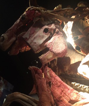
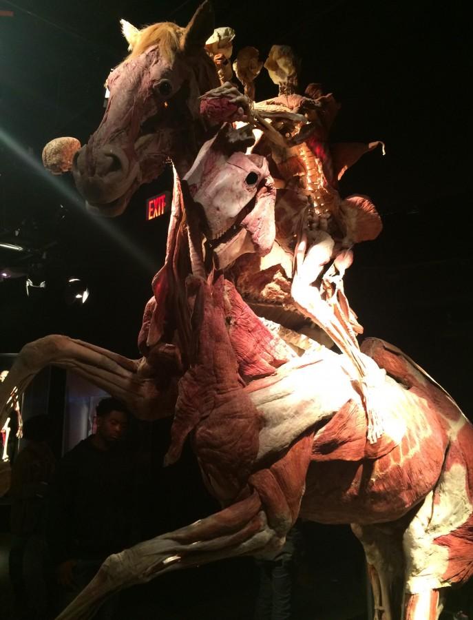
Other displays they saw included a showcase of all of the body’s blood vessels, from the brain to the toe, and two people riding on a horse with their bodies opened up for observations. Both exhibits showed how human muscles work in our everyday exercises, while maintaining an artistic touch.
“It was a mixture of something very informative and very artistic. Everything was displayed in a way that you could see everything and the aesthetic value of it,” senior Kyle Arroyo said.
The trip received such positive feedback from students that Dr. Ponticello has decided to continue it for another year. She reasoned that textbooks are not enough, and looking at real, preserved bodies gives students a learning experience unattainable anywhere else.
“I thought it was very cool because people often take for granted about what happens in the body. This exhibit really allows you to see the functional processes in the body,” Angus said.


